The Good, The Bad and The Ugly!
They can be mean, destroying a pretty face or leg.
They can be ugly, growing into large masses that disfigure.
They can be expensive.
They can be very difficult to treat.
What are they? They are sarcoids, the most common type of skin tumor in horses.
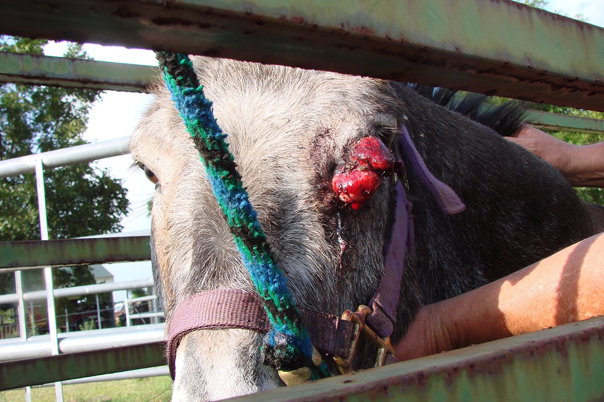
Sarcoids are more common in young horses but they can occur in any horse of any age. Sarcoids can occur in any equid including in donkeys and zebras.
Sarcoids are caused by the Bovine Papilloma Virus (Cattle Wart Virus). They are believed to enter the body through wounds or breaks in the skin. Even the small hole of an insect bite can be an entry point.
Sarcoids are commonly found in the ears (a preferred feeding site of biting gnats), around the eyes where insects feed on tears, on the lower legs where injuries are most common, in the axilla (armpit region) and in the groin. Sarcoids do not metastasize (spread to internal organs) but they can be locally aggressive. Sarcoids can become very large in size and can spread covering an increasingly larger area.
There are 5 types of Sarcoids.
1. Occult
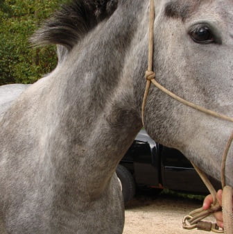
These sarcoids are typically flat, oval to circular areas of hair loss skin The skin still has some hair and small bumps can be felt through the skin. They may resemble ringworm or rub marks from tack. They are commonly seen on the nose and side of the face, the axilla (armpit) and groin. If accidently traumatized these sarcoids have the potential to develop into a more serious type of sarcoid. In this horse the skin lesion is barely visible from a distance at the base of the neck. However, a closer look shows an oval area where the hair is thinner. The small bumps can be easily felt in the skin.
.
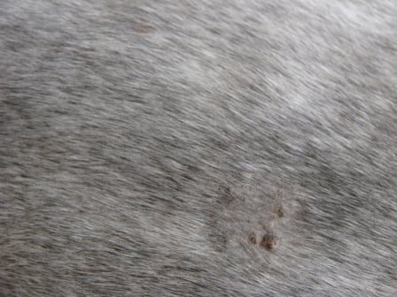
2. Verrucous
These sarcoids look a lot like warts, they tend to be gray in color with no hair overlying. Verrucous sarcoids may flake off in thin layers or be smooth and shiny. They may be single lesions or in clusters of sarcoids.
In this photo you can see 3 different types of sarcoids. the top red, raised bump is a nodular sarcoid that is undergoing fibroblastic transformation. The calipers are placed over a nodular sarcoid. To the left of the calipers is a large field of verrucous sarcoids that look like raised scaly skin.

3. Nodular
This form appears as firm, raised, round nodules. They can appear anywhere on the body but are often seen in the axilla (armpit), on the inside of the thigh and groin and under the skin of the eyelids. They may be singular or in groups. They may be covered by normal skin or covered with ulcerated skin. They are usually firmly attached to the overlying skin.
4. Fibroblastic
These are red fleshy masses that grow quickly, bleed easily and have ulcerated surfaces. They look like exuberant granulation tissue (Proud Flesh) and can develop at the site of previous wounds. They can be found anywhere on a horses body. They usually start as a smaller less aggressive form of sarcoid that become aggressive following trauma, biopsy or treatment.

5. Mixed Sarcoid
In these cases the lesion shows the qualities of two or more sarcoid groups.
Some Important Sarcoid Facts
Sarcoids are common; geldings appear more frequently affected.
- All equid species are susceptible even donkeys and zebras.
- Although sarcoids are a type of tumor (cancer) they do not metastasize (spread to internal organs).
- Once a sarcoid horse, always a sarcoid horse! Once the virus has entered the horses’ body it is at greater risk to develop more sarcoids.
- Sarcoids can develop anywhere on the horse’s skin, but more common sites include the chest, groin, sheath and face (especially around the eyes and mouth).
- Trauma of any nature to a sarcoid is likely to aggravate it.
- No two sarcoids are the same; each sarcoid needs to be assessed on an individual basis.
- Sarcoids can be unpredictable in all aspects of their development and treatment and the way they respond to treatment. It is this variability that makes sarcoids such a challenge for both owners and veterinarians.
Diagnosis of Skin Lesions
Sarcoids can look a lot like other types of skin lesions. the ONLY definitive way to diagnose a sarcoid is through biopsy. the tissue is submitted to a pathologist who examines the tissue under a microscope and confirms the diagnosis.
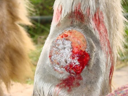
This horse has a red, raised mass Aluspray (a silver spray), has been previously applied. A biopsy confirmed this lesion as Habronemiasis, not a sarcoid.
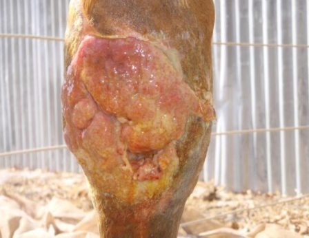
This horse had a wound, the wound has filled with tissue that is red, raised and bumpy in appearance. A biopsy confirmed this lesion as exuberant granulation tissue or “Proud Flesh”.
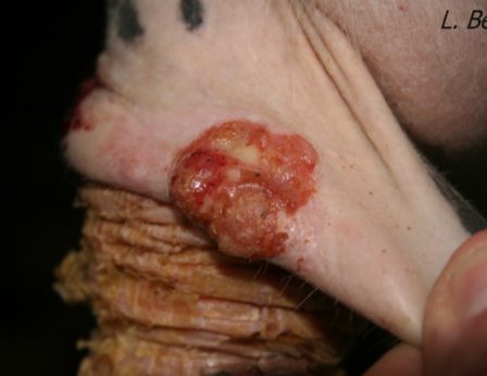
This horse has a red, raised bumpy mass on his sheath. A biopsy confirmed this to be a case of squamous cell carcinoma, a type of cancer.
As you see the outward appearance of skin lesions can be confusing. In some cases a biopsy is required to make a diagnosis so that the correct treatment is given. The wrong treatment can be just as harmful as no treatment at all. Once we have a diagnosis it’s time to decide what action is best. In many cases it is a combination of therapies.
Sarcoid Treatment
Unfortunately there isn’t a magical cure-all treatment for sarcoids! Apparently, there are over 40 different sarcoid treatments world-wide which clearly demonstrates that there is no one single method that will be effective in each and every case! Horses should be treated at an early stage in the disease when lesions are small and treatment before 4 years of age appears to have a better prognosis.
Each and every sarcoid is different; they are unpredictable by nature and no matter how similar two sarcoids look, a treatment that works for one might not work for another. It is extremely important to remember that each sarcoid needs to be assessed by a veterinarian on an individual basis before any treatment is started. Inappropriate treatment can easily convert a simple sarcoid into something very nasty, very quickly
Sarcoid Treatment Options
- Benign neglect and monitoring – Small “Occult” sarcoids may be better left alone and left undisturbed. Treatment and biopsy can make many sarcoids worse. The sarcoid should be measured and photographed. If the lesion becomes aggressive an appropriate treatment course can begin.
- Surgical excision – Surgery can be very helpful with small lesions or lesions where a large margin of tissue can be removed without causing undue scarring. Failure to remove an adequate margin frequently results in recurrence. In some cases surgical removal is done in conjunction with post-operative chemotherapy, immunotherapy or radiation.
- Laser surgery – A second surgical technique involves the use of a surgical laser. Laser surgery has the advantage of hemostasis (bleeding control). Because laser energy is absorbed by the surrounding tissues, tumor cells are killed up to 2mm around the wound edges. The downside is the potential to aerosolize infectious particles from the tissue being removed, the consequences to the horse and surgeon are undetermined. Most cases combine laser removal with another form of treatment such as chemotherapy or immunotherapy.
- Cryosurgery – Liquid nitrogen is used to freeze the tissue, the frozen tissue dies and sloughs off. The tissue being treated cannot be very thick or the deeper tissues will not freeze adequately. Generally this is done on small lesions or in larger lesions after the tissue has been surgically debulked.
- Hyperthermia – In this technique a specialized device is used to deliver heat into the tissue with a special probe, the heat destroys the tumor cells and the tissue subsequently dies. Hyperthermia units are not readily available in equine practice.
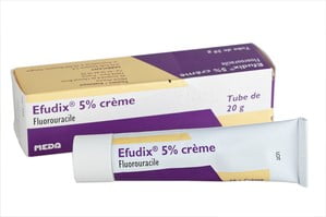
- Chemotherapy – Two drugs are currently available, 5 Fluorouracil and Cisplatin.
5 Fluorouracil or 5 FU is a topical chemotherapy treatment. Four treatments are given with the 1st and 2nd treatments 24 hours apart followed by a 3rd treatment 48 hours later and a 4th treatment 48 hours that. The sarcoid tissue will become swollen and inflamed and may look worse before it looks better. Local swelling can be painful to the horse and an anti-inflammatory will be prescribed. This drug has been used extensively in Europe with excellent results.
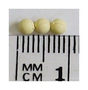
Cisplatin – A chemotherapy drug that is available in two forms. An injectable solution is available that must be mixed with sterile oil to give it a slow release property. A set of 3-5 injections are done every 2-3 weeks. Local swelling should be expected. A second formulation of cisplatin is a bead. The beads have been impregnated with cisplatin. The beads are inserted into the tissue using tiny incisions and a single suture. The beads slowly release the drug into the tissue over a 30 days period. Treatment can be repeated as necessary.
- Immunological stimulation – Bacillus Calmette-Guerin (BCG) injection works well for nodular and fibroblastic lesions around the eyes but is much less effective elsewhere and should not be used for sarcoids on the limbs as these may become much worse. The product is injected 3 times at weekly intervals. Serious allergic reactions can occur in some horses.
- Autologous vaccination – In this treatment an existing sarcoid mass is removed and then frozen in liquid nitrogen. The frozen mass is then diced into small bits and inserted back into the horse in a series of subcutaneous pockets under the mane. Significant tissue reaction will occur at the implantation sites. Response varies from case to case. Some horse achieve full remission while others do not respond.
- Radiation – By far the gold standard in treatment options is radiation. Radiation has the advantage that no tissue is lost with minimal scarring and distortion. The major disadvantage to radiation involves cost, general anesthesia and patient transport. Radiation is only available in a university hospital and the horse must be under general anesthesia to be placed near the machine that delivers the radiation. Multiple treatments are done depending upon the size of the area involved and the tissue response.
- Antiviral Medication – Two drugs are currently available.
Immiquimod (Aldara), a human immune response modifier with potent antiviral and antitumor activity that is used to treat skin cancer and genital warts in humans. The cream is applied over the sarcoid three times weekly. Local swelling and reaction will occur and many sarcoids look worse before they get better. Treatment may take 3-4 months. The advantage is that owners can apply the cream themselves.
Acyclovir is an antiviral drug available in a cream or ointment formulation. The cream is applied daily for 2-6 months. - Bloodroot Extract – Sold under the trade name of Xxterra, this is an economical option for smaller sarcoids and sarcoids that are not near the eye. The cream is applied daily for 4-5 days and the tissue dies and sloughs off. The area becomes painful during this process and some horse will require sedation to make the treatment applications.
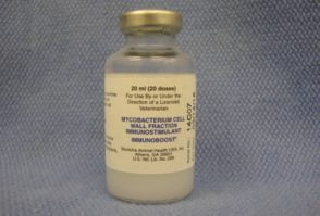
Bacillus Calmette-Guerin (BCG) injection
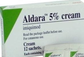
Immiquimod (Aldara)
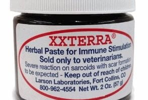
Bloodroot Extract
Treatment of sarcoids can be complicated. No two sarcoids respond the same. Ultimately the best treatment option depends on a combination of the type of sarcoid, the location or the sarcoid (s), financial constraints and compliance of the horse and owner. In many cases more than one type of therapy will be needed and treatment can be prolonged.
